Home » Without Label » Muscles Of The Chest Abdomen : How To Build Huge Chest / In this video we will go over the main muscles in the chest, abdomen, pelvis and back.
Muscles Of The Chest Abdomen : How To Build Huge Chest / In this video we will go over the main muscles in the chest, abdomen, pelvis and back.
Muscles Of The Chest Abdomen : How To Build Huge Chest / In this video we will go over the main muscles in the chest, abdomen, pelvis and back.. If you've pulled a muscle—particularly in your chest, abdomen, or upper/middle back area—you may experience chest tightness and pain when you engage in activities. There is a printable worksheet available for download here so you can take the quiz with pen and paper. This complex structure consists of numerous layers, from superficial to deep: In the rear of the abdomen are the back muscles and. In front of the fascia are the abdominal muscles and skin.
One of the main smooth muscles inside the chest is the diaphragm. #anatomy of the chest and stomach. Start studying muscles of the chest and abdomen. The muscles of the back are in 5 layers, one beneath another. The muscles of the abdomen, lower back, and pelvis are separated from those of the chest by the muscular wall of the diaphragm, the critical breathing muscle.
Anterior chest and abdomen from www.purposegames.com Moving down the trunk of the cat from the chest to the abdomen, i was able to identify the latissimus dorsi, internal oblique, transverse abdominus, rectus abdominus, linea alba, and external oblique. This may also radiate to the shoulder, arm, or even cause abdominal discomfort. The abdominal wall can be divided into two sections: Start studying muscles of the chest and abdomen. Muscles the dominant muscle in the upper chest is the pectoralis major. Anatomy of the chest and stomach, human anatomy, anatomy of the chest and stomach. The muscles of the back are in 5 layers, one beneath another. #anatomy of the chest and stomach.
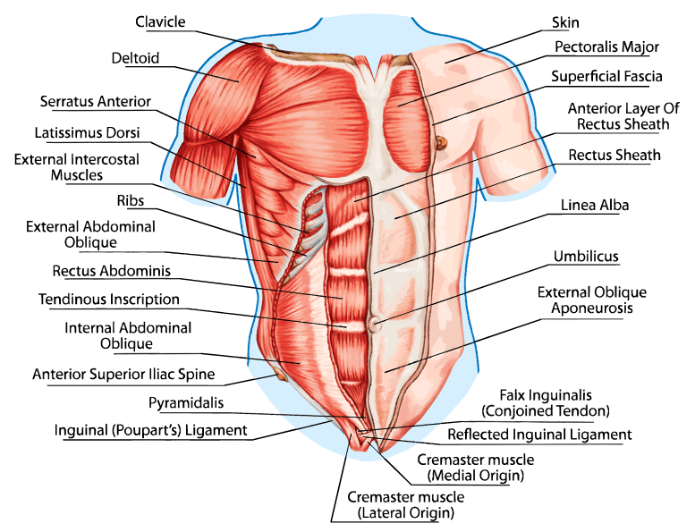
The rectus abdominis is positioned between the ribs and the pubic bone at the front of the pelvis, and is actually made up of 8 distinct muscle bellies.
In the front, the abdomen is protected by a thin, tough layer of tissue called fascia. This complex structure consists of numerous layers, from superficial to deep: A diaphragm spasm is an involuntary contraction of the muscle that divides the upper abdomen and chest. Moving down the trunk of the cat from the chest to the abdomen, i was able to identify the latissimus dorsi, internal oblique, transverse abdominus, rectus abdominus, linea alba, and external oblique. Muscles of the chest and abdomen. The external obliques, the internal obliques, the transversus abdominis, and the rectus abdominis (figure 1, figure 2, and table 1). If you've pulled a muscle—particularly in your chest, abdomen, or upper/middle back area—you may experience chest tightness and pain when you engage in activities. One of the most common symptoms of pulling a chest muscle is pain around the affected muscle. Muscle diagram free weights muscle anatomy chest muscles nursing tips medical science upper body back pain scorpio. Originates from the upper portion of the sternum. Inserts along almost the entire length of the humerus and on the fascia covering the proximal end of the forearm. This lesson covers the following objectives: Rectus abdominis and external oblique from a muscular view, the ventral abdomen consists of the rectus abdominis and the pyramidal muscles.
#anatomy of the chest and stomach. The abdominal wall can be divided into two sections: In the rear of the abdomen are the back muscles and. If you've pulled a muscle—particularly in your chest, abdomen, or upper/middle back area—you may experience chest tightness and pain when you engage in activities. This lesson covers the following objectives:
Chest - wikidoc from www.wikidoc.org Back muscles chart 12 photos of the back muscles chart back muscles chart, back muscles diagram and ligaments, back muscles diagram lats, back muscles diagram massage, upper back muscles chart, human muscles, back muscles chart, back muscles diagram and ligaments, back muscles diagram lats, back muscles diagram massage. Layers of the abdominal wall. The abdominal head of the pectoralis major muscle is one of three origins for the pectoralis major. There are four pairs of abdominal muscles that cover the anterior and lateral abdominal region and meet at the anterior midline. In the front, the abdomen is protected by a thin, tough layer of tissue called fascia. Moving down the trunk of the cat from the chest to the abdomen, i was able to identify the latissimus dorsi, internal oblique, transverse abdominus, rectus abdominus, linea alba, and external oblique. View muscles of the chest, abdomen and thigh (deep dissection, 1 of 2).jpg from biology misc at miami dade college, miami. If you've pulled a muscle—particularly in your chest, abdomen, or upper/middle back area—you may experience chest tightness and pain when you engage in activities.
This complex structure consists of numerous layers, from superficial to deep:
A diaphragm spasm is an involuntary contraction of the muscle that divides the upper abdomen and chest. One of the main smooth muscles inside the chest is the diaphragm. The two largest and most superficial are the trapezius and the latissimus dorsi. It arises from the fascia of the external oblique muscle. In some cases, the strain may be severe enough to cause pain when breathing. But it also could be the sign of gerd, a peptic ulcer, or something very serious, such as heart attack or pulmonary embolism. In the front, the abdomen is protected by a thin, tough layer of tissue called fascia. Skin, superficial fascia, muscles and their respective fasciae, and peritoneum. In front of the fascia are the abdominal muscles and skin. Learn vocabulary, terms, and more with flashcards, games, and other study tools. The muscles of the abdomen, lower back, and pelvis are separated from those of the chest by the muscular wall of the diaphragm, the critical breathing muscle. William blahd on webmd says that pulled muscle, strains, and tears can damage the muscle fibers and tendons. Using an engaging and simplified teaching style, our expert.
In the front, the abdomen is protected by a thin, tough layer of tissue called fascia. Using an engaging and simplified teaching style, our expert. Signs and symptoms of pulled chest muscles. View muscles of the chest, abdomen and thigh (deep dissection, 1 of 2).jpg from biology misc at miami dade college, miami. In front of the fascia are the abdominal muscles and skin.
How To Open Up The Chest Muscles To Prevent Forward ... from livelovefruit.com A diaphragm spasm is an involuntary contraction of the muscle that divides the upper abdomen and chest. Moving down the trunk of the cat from the chest to the abdomen, i was able to identify the latissimus dorsi, internal oblique, transverse abdominus, rectus abdominus, linea alba, and external oblique. 12 photos of the anatomy of the chest and stomach. Back muscles chart 12 photos of the back muscles chart back muscles chart, back muscles diagram and ligaments, back muscles diagram lats, back muscles diagram massage, upper back muscles chart, human muscles, back muscles chart, back muscles diagram and ligaments, back muscles diagram lats, back muscles diagram massage. Signs and symptoms of pulled chest muscles. This complex structure consists of numerous layers, from superficial to deep: Chest pain, chest tightness, chest pressure, palpitations. Start studying muscles of the chest and abdomen.
This is an online quiz called muscles of the chest and abdomen labeling.
The external obliques, the internal obliques, the transversus abdominis, and the rectus abdominis (figure 1, figure 2, and table 1). This is an online quiz called muscles of the chest and abdomen labeling. In some cases, the strain may be severe enough to cause pain when breathing. The muscles of the abdomen, lower back, and pelvis are separated from those of the chest by the muscular wall of the diaphragm, the critical breathing muscle. One of the most common symptoms of pulling a chest muscle is pain around the affected muscle. Moving down the trunk of the cat from the chest to the abdomen, i was able to identify the latissimus dorsi, internal oblique, transverse abdominus, rectus abdominus, linea alba, and external oblique. A diaphragm spasm is an involuntary contraction of the muscle that divides the upper abdomen and chest. The abdominal wall can be divided into two sections: Inserts along almost the entire length of the humerus and on the fascia covering the proximal end of the forearm. The rectus abdominis is positioned between the ribs and the pubic bone at the front of the pelvis, and is actually made up of 8 distinct muscle bellies. There is a printable worksheet available for download here so you can take the quiz with pen and paper. Chest and abdominal pain may be caused by something as simple as gas. #anatomy of the chest and stomach.